
Figure 7.
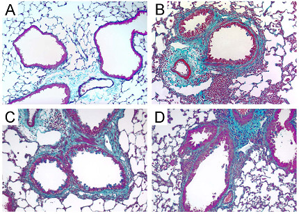
Increased airway subepithelial collagen deposition after treatment with Aspergillus antigen. Representative lung sections from PBS-treated mice show minimal trichrome staining around small airways (A) (similar findings from Mcp-1-/- and Ccr2-/- control mice are not shown). Increased trichrome staining is noted around small airways in Aspergillus antigen-treated wild-type (B), Mcp-1-/- (C) and Ccr2-/- (D) mice. Blue staining around airways represents collagen. Aspergillus antigen exposure and sample collection are described in methods. Magnification, 20× objective.