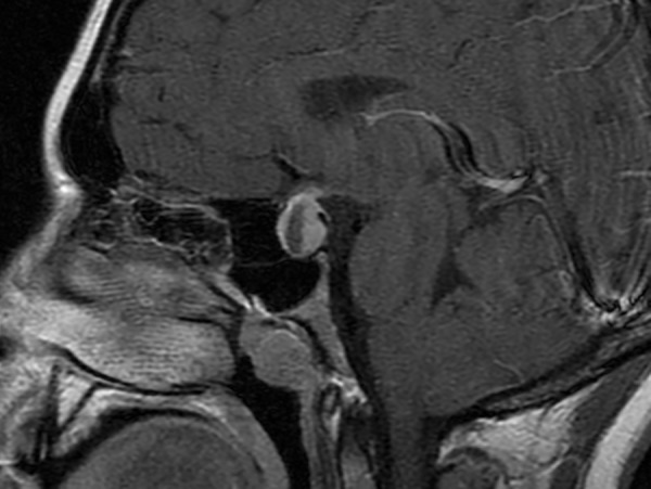
Figure 1. Neuroimaging of Pituitary Mass.

T1 MRI with contrast showing a 2 cm ring-enhancing suprasellar mass with effacement of the optic chiasm.

T1 MRI with contrast showing a 2 cm ring-enhancing suprasellar mass with effacement of the optic chiasm.