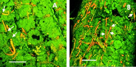
FIG. 5.
Micrographs of field samples of Chara viewed under the confocal scanning laser microscope. Microcolonies of unicellular and filamentous cyanobacteria were present on the surfaces of internodes (A) and verticiles (B) of Chara. The arrows indicate heterocysts. (A) Scale bar = 100 μm. (B) Scale bar = 70 μm.

