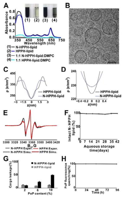
Figure 3.
N-HPPH-lipid bilayers with enhanced hydration. A) Optical absorption of hydrated PoP lipid films shortly after water addition. B) Representative cryo-electron micrographs showing structures formed by N-HPPH-lipid at a 1:1 DMPC:PoP molar ratio. 40 nm scale bar is shown. C), D) Bilayer electron density determined by X-ray diffraction in bilayers formed with a 1:1 DMPC:PoP molar ratio. E) Electron spin resonance spectra showing differences in lipid ordering between N-HPPH-lipid and HPPH-lipid bilayers (containing DMPC and PoP and probed with 0.5 molar % 10 PC spin label). F) Hydrolysis of N-HPPH-lipid liposomes during aqueous storage. G) Sulforhodamine B leakage over 24 hr in saline, in liposomes containing indicated molar percentage of PoP, together with 35 mol. % Chol and the remaining composition of DMPC. H) Structural intactness of cargo-loaded, N-HPPH-liposomes (PoP:DMPC:Chol; 10:35:55 molar ratio) based on fluorescence self-quenching. Data show mean ± s.d. for n=3.

