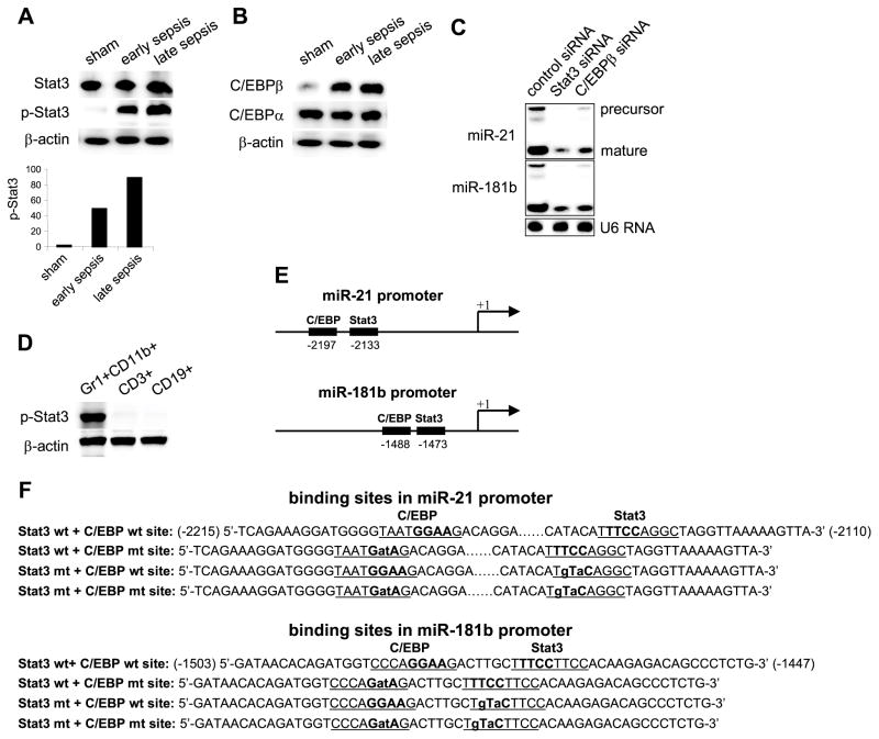
Figure 1. Stat3 and C/EBPβ activate miR-21 and miR-181b expression in sepsis Gr1+CD11b+ MDSCs.
Sepsis was induced by cecal ligation and puncture (CLP). (A and B) Bone marrow cells were harvested from septic mice that were moribund and sacrificed at days 1–5 (representing early sepsis) and at days 6–28 (representing late sepsis) as well as sham mice. Gr1+CD11b+ cells were then purified using magnetic beads and pooled from 6 mice per group. Cell lysates were prepared and levels of total and phosphorylated (p-Stat3; Tyr705) Stat3 (A), and C/EBPβ proteins (B) were determined by immunoblot. The results are representative of two experiments. Lower panel in A shows densitometry of the p-Stat3 bands. Values were normalized to β-actin and are presented relative to sham, which is set at 1-fold. (C) Knockdown of Stat3 or C/EBPβ in sepsis MDSCs inhibits miR-21 and miR-181b expression. Gr1+ CD11b+ cells were isolated from the bone marrow of late septic mice, pooled (n = 6 mice per group), and transfected with Stat3-specific, C/EBPβ-specific, or control siRNA. After 36 hr in culture, cells were harvested and levels of miR-21 and miR-181 were determined by northern blot. Levels of the U6 RNA were also measured as an internal control. The knockdown was confirmed by western blotting (not shown). (D) Phosphorylation of Stat3 in the bone marrow during sepsis is restricted to the Gr1+CD11b+ MDSCs. CD3+ T cells were isolated from whole bone marrow cells by positive selection using anti-CD3 magnetic beads. To isolate CD19+ cells, whole bone marrow cells from late septic mice were first depleted of Gr1−CD11b+ cells (which mostly consists of CD19+ and CD11c+ cells). CD19+ cells were then positively selected from the depleted cell population using anti-CD19 magnetic beads. The results are representative of two immunoblots using two different cell preparations. (E) A schematic diagram depicting the Stat3 and C/EBPβ binding sites in the miR-21 and miR-181b promoters. (F) The miR-21 and miR-181b promoter fragments used in the luciferase gene constructs (see Fig. 6). The Stat3 and C/EBP binding sites are underlined. The core nucleotides of the consensus binding sites are bolded. Lower case letters indicate the mutation introduced in the binding site.

