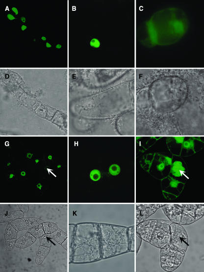
Figure 1.
Localization of GFP:CycD1 Fusion Protein and GFP in Living Cells.
(A) to (F) Transiently expressed in Arabidopsis cell culture.
(A) and (B) Confocal images of GFP fluorescence in cells transformed by GFP-CycD1;.
(C) and (F) Confocal image of soluble GFP fluorescence and corresponding phase contrast image.
(D) and (E) Corresponding phase contrast images.
(G) to (L) Stable expression in BY-2 cell culture. Arrows indicate mitotic cells.
(G) and (H) Confocal images of GFP fluorescence in cells transformed by GFP-CycD1;.
(I) and (L) Confocal image of soluble GFP fluorescence and corresponding phase contrast image.
(J) and (K) Corresponding phase contrast images.

