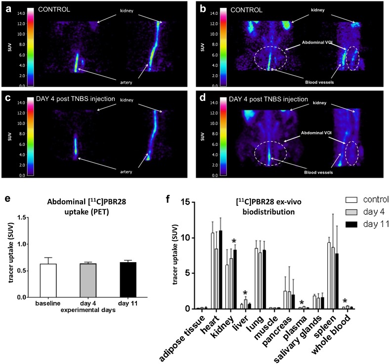
Fig. 3.
PET imaging and ex vivo biodistribution of peripheral organs and tissues as a function of disease progression. Healthy rats or TNBS-treated rats at either day 4 or day 11 post-TNBS injection were injected intravenously with [11C]PBR28 (29 ± 11 MBq) and subjected to the a 60-min PET scan of the abdomen under isoflurane anesthesia: examples of sagittal and coronal sections showing the a first 50 s of the scan and b the last 10 min of the scan of a control rat; c the first 50 s of the scan and d the last 10 min of the scan of a rat 4 days after TNBS injection; e abdominal uptake (SUV) of [11C]PBR28 [mean ± standard deviation] in TNBS-treated and control rats. Immediately after the PET scan (65 min after tracer injection), animals were euthanized by cardiac puncture under deep sevoflurane anesthesia. Organs and tissues were harvested for ex vivo biodistribution. [11C]PBR28 uptake in major organs and tissues as determined by f ex vivo biodistribution (mean ± standard deviation). Statistically significant differences between TNBS-treated animals and the control group are indicated with an asterisk: *p < 0.05 (ANOVA with Dunnett post hoc test).

