Fig. 5.
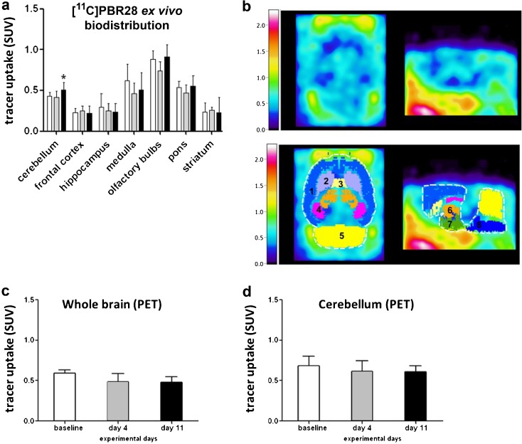
Ex vivo biodistribution and PET imaging of the brain. a Ex vivo biodistribution of [11C]PRB28 (65 min) in the brains of control rats (n = 6) and TNBS-treated rats sacrificed at day 4 (n = 6) and day 11 (n = 6). b An example of a sagittal and coronal [11C]PRB28 PET image of the brain of a rat subjected to TNBS treatment (day 4) and its overlay with a MRI template with selected regions, as indicated by different colors and the numbers: 1 = cortex, 2 = caudate putamen, 3 = septum, 4 = hippocampus, 5 = cerebellum, 6 = thalamus, 7 = hypothalamus, 8 = pons + medulla. [11C]PRB28 uptake (50–60 min) in c whole brain and d cerebellum was measured by PET imaging. Data are presented as mean ± standard deviation. Statistically significant differences between TNBS-treated animals and the control group are indicated with an asterisk: *p < 0.05 (ANOVA with Dunett post hoc test).

