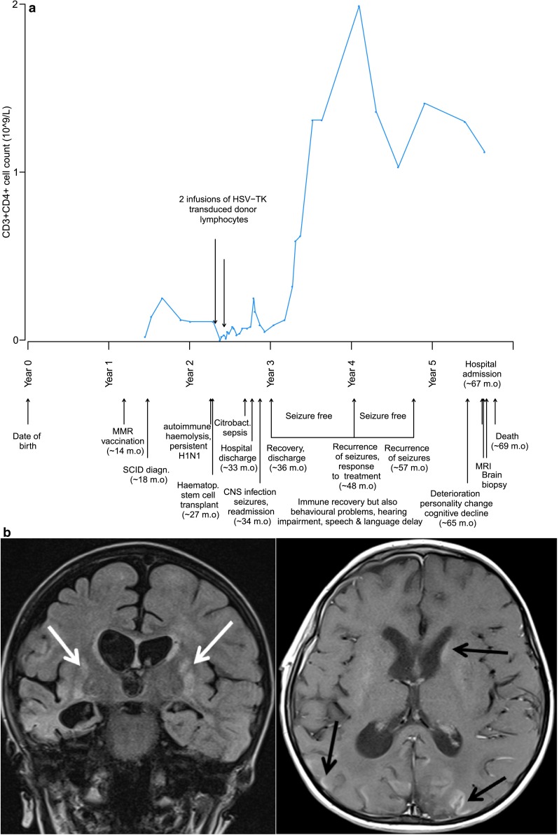
Fig. 1.

a Patient timeline presenting important clinical events (m.o. = months old) and immune recovery of CD3+ CD4+ T cells over this period. b MRI brain scan showing coronal T2-weighted FLAIR (FLuid Attenuated Inversion Recovery) and axial post-contrast T1-weighted images with bilateral basal ganglia lesions (white arrows) and enhancing cortical and deep grey matter lesions (black arrows). The pattern was typical of those described in subacute panencephalitis
