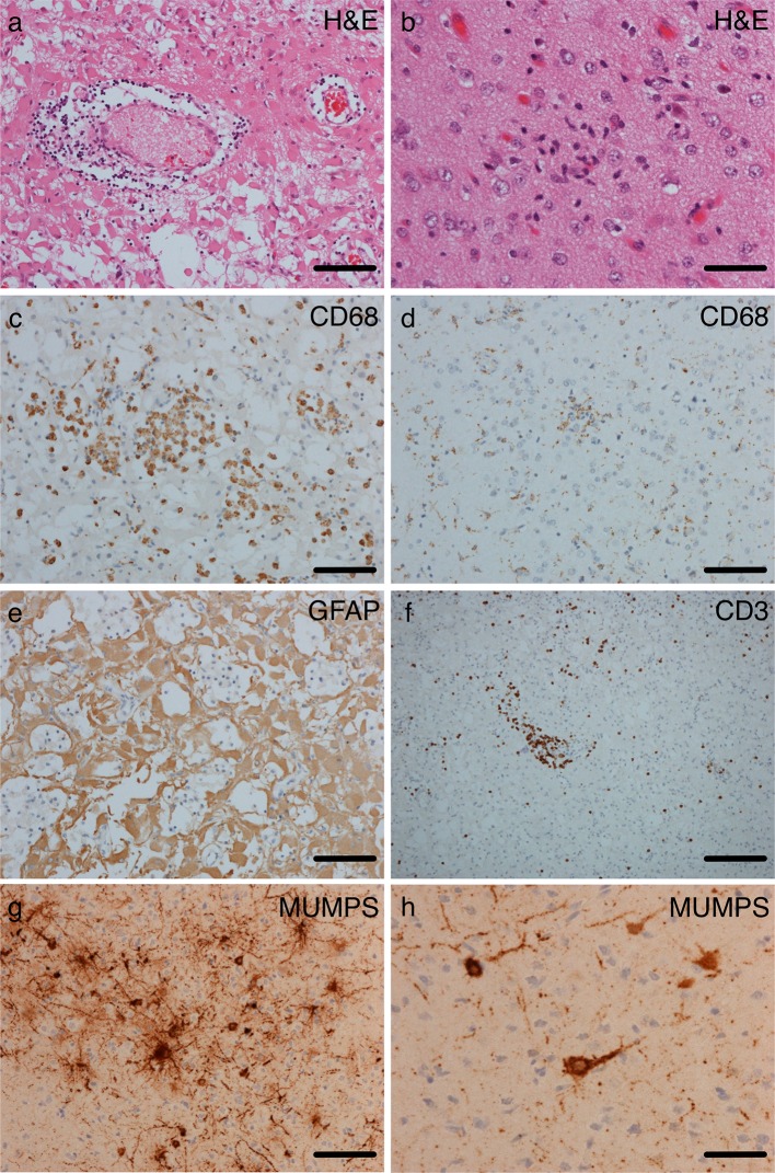
Fig. 2.
Much of the cortex showed significant tissue damage with neuronal loss. There was prominent reactive gliosis composed of plump astrocytosis with abundant eosinophilic cytoplasm and immunoreactivity for glial fibrillary acidic protein (GFAP) (a, e) with relative sparing of the superficial layers of the cortex. Some areas showed perivascular lymphocytosis and collections of macrophages (a, c). In others, with better neuronal preservation, there were foci of microglial nodules, neuronophagia, generalised microgliosis (demonstrated on CD68 staining b, d)) and patchy parenchymal and perivascular lymphocytes, the majority of which were positive for the T cell markers (f). Occasional foci of mineralisation were observed, but there was no vasculitis and no viral inclusions. The pathological changes extended into the underlying white matter (data not shown), which showed patchy loss of myelin staining on luxol fast blue. There was focal chronic inflammation in the leptomeninges. Mumps immunohistochemistry was positive in a neuronal pattern (g, h). We observed no specific staining for other pathogens HSV1 or 2, CMV, EBV, toxoplasma, JC virus, bacteria (Gram and Gram Twort), acid-fast bacteria (Ziehl-Neelson) or fungi (Grocott), and no evidence of intracellular or intranuclear viral inclusion bodies. a, b Haematoxylin and eosin (H&E), c, d CD68 immunohistochemistry, e GFAP immunohistochemistry, f CD3 immunohistochemistry. Scale bars-b, h 50 μm, a, c, d, e, g 100 μm, f 200 μm

