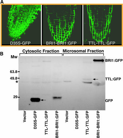
Figure 4.
TTL Is a Membrane-Associated Protein.
(A) Confocal microscopic analysis of GFP fusion proteins in the root tips of 4-d-old transgenic plants containing the BRI1-BRI1:GFP or TTL-TTL:GFP transgene. The green fluorescent signal of the GFP itself driven by two copies of the strong 35S promoter of Cauliflower mosaic virus was used as a control for cytosolic and nuclear localization.
(B) Subcellular fractionation analysis of the TTL localization. Cytosolic (lanes 1 to 4) and membrane fractions (lanes 5 to 8) were prepared from the 7-d-old seedlings of transgenic plants containing the pPZP212 vector alone, D35S-GFP, TTL-TTL:GFP, or BRI1-BRI1:GFP transgene. The similar amount of total proteins of each fractionation was analyzed for the presence of GFP or GFP-fusion proteins by immunostaining with anti-GFP antibodies (Molecular Probes, Eugene, OR). The asterisk indicates a nonspecific cross-reacting band, and the arrow indicates the TTL:GFP fusion protein.

