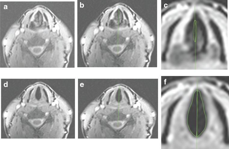
Fig. 5.
a, d: selected images showing glottal area during phonation (a) and respiration (d); b, e: A vertical line was drawn from anterior commissure bisecting the posterior commissure and the cervical vertebra dividing the triangular larynx into left and right glottal areas; c, f: Regions of interest (ROI) drawn to measure left and right phonation (c) and respiration (f) areas

