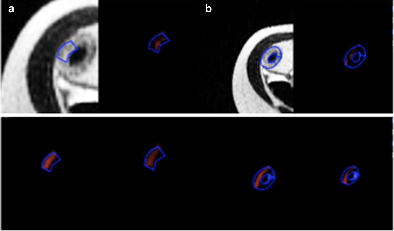
Fig. 1.
Examples of small (a) and large (b) regions of interest drawn on axial half-Fourier RARE sequence images in diseased bowel subsequently resected along with the texture maps at fine (upper right panel), medium (lower left panel), and coarse (lower right panel) texture scales. Small ROI = 42 pixels. Large ROI = 169 pixels

