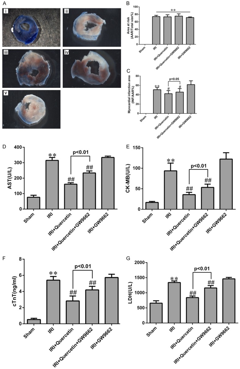
Figure 2.

Effects of quercetin and PPARγ inhibitor on myocardial infarct size and myocardial enzyme leakage. A. Myocardial infarct size was determined by TTC staining. Representative TTC-stained myocardial sections were shown. Blue-stained areas indicate normal tissue, red-stained areas indicate AAR and unstained pale areas indicate INF. AAR and INF sizes were subsequently measured. (i) Sham-operated mice treated with vehicles (Sham). (ii) Myocardial IRI mice treated with vehicles (IRI). (iii) Myocardial IRI mice treated with quercetin (IRI+Quercetin). (iv) Myocardial IRI mice treated with GW9662 and quercetin (IRI+Quercetin+GW9662). (v) Myocardial IRI mice treated with GW9662 (IRI+GW9662). B. The percentage of AAR to the total left ventricular area 24 h post IRI in the indicated groups. C. The percentage of INF area to AAR 24 h post IRI in the indicated groups. D-G. Total AST, CK-MB, cTnT and LDH levels in arterial blood (n=6). **P<0.01 vs Sham group, #P<0.05, ##P<0.01 vs IRI group. The experiment was repeated six times.
