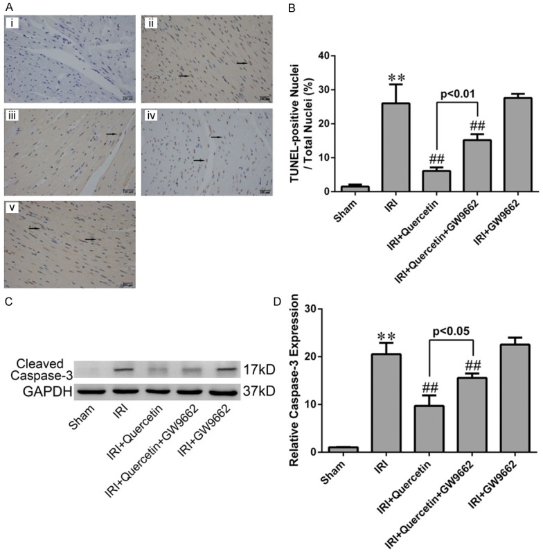
Figure 7.

Effects of quercetin and PPARγ inhibitor on cardiomyocyte apoptosis in vivo. (A) TUNEL staining for left ventricular tissue from different treatment groups. Nuclei with brown staining indicated TUNEL-positive cells (black arrows) and blue staining indicate TUNEL-negative cells (magnication ×400). (i) Sham-operation mice treated with vehicles (Sham). (ii) Myocardial IRI mice treated with vehicles (IRI). (iii) Myocardial IRI mice treated with quercetin (IRI+quercetin). (iv) Myocardial IRI mice treated with GW9662 and quercetin (IRI+quercetin+GW9662). (v) Myocardial IRI mice treated with GW9662 (IRI+GW9662). (B) Quatification of apoptotic rate based on TUNEL staining (five fields for each specimen). (C) Western blot analysis of cleaved caspase-3 in the indicated groups. Densitometry was shown in (D). The values were presented as mean ± SD. **P<0.01 vs Sham group; ##P<0.01 vs IRI group. All experiments were repeated six times.
