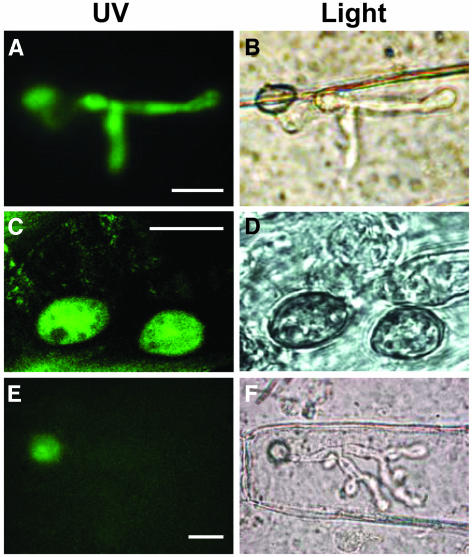
Figure 5.
Localization of Ace1-GFP Fusion Protein in M. grisea Appressoria.
(A) GFP fluorescence of appressoria and infectious hypha after penetration of barley leaves. Conidia of transformant IF22 expressing GFP under the control of the ACE1 promoter were inoculated on detached barley leaves and incubated at 26°C. At 36 h after infection, epidermal strips were removed from the infected leaf and observed under a microscope. Under blue light, GFP fluorescence is detected both in appressorium and in infectious hyphae. Bar = 10 μm.
(B) Same view as (A) under bright field. An appressorium and its infectious hypha located in the underlying epidermal cell are visible.
(C) Ace1-GFP fluorescence of appressoria on the surface of barley leaves. Conidia of transformant HB41 expressing the Ace1-GFP fusion protein under the control of ACE1 promoter were inoculated on detached barley leaves incubated for 24 h at 26°C and observed with confocal laser scanning microscopy. Under blue light, fluorescence of the Ace1-GFP fusion protein is detected exclusively in the cytoplasm of the appressoria. Bar = 10 μm.
(D) Same view as (C) under bright field. Two germinated spores and appressoria are visible on the leaf surface.
(E) Ace1-GFP fluorescence of appressoria after penetration of barley leaves. Conidia of transformant HB44 expressing the Ace1-GFP fusion protein were inoculated on detached barley leaves and incubated at 26°C. Epidermal strips were removed from the infected leaf 40 h after inoculation and observed under a microscope. Under blue light, GFP fluorescence is detected in the appressorium but not in infectious hypha. Bar = 10 μm.
(F) Same view as (E) under bright field. An appressorium and its infectious hypha located in the underlying epidermal cell are visible.

