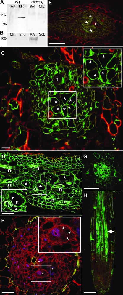
Figure 3.
Immunolocalization of AtFH6 and Actin in Galls and Root Apex.
(A) and (B) Cell fractionation and protein gel blot analyses of wild-type or homozygous csq2/csq2 plants.
(A) Mouse antibody AbAFH6 was used to detect the protein in soluble (Sol) and microsomal (Mic) fractions.
(B) The purified plasma membrane fraction (P.M.) of Arabidopsis wild-type cells contained a higher amount of AtFH6 than the microsomal fraction (Mic) or the endomembranes (End). No signal was detected in the cytosolic fraction (Sol).
(C) and (D) Immunolocalization of the AtFH6 protein in gall sections 5 (C) and 7 dpi (D) of wild-type plants. Fluorescent signal is detected in the plasma membrane (arrowheads) of giant cells (asterisks) and surrounding cells. n, nematodes.
(E) Immunolocalization of the AtFH6 protein in a gall section 7 dpi of csq2/csq2 plants showing no fluorescence.
(F) Immunofluorescence detection of actin in galls 5 dpi of wild-type plants. A detail of a giant cell (asterisks) showed actin localization in the cell cortex (arrowheads) and less within the cytoplasm. Actin is visualized in red and 4′,6-diamidino-2-phenylindole (DAPI)-stained nuclei in blue.
(G) and (H) Immunolocalization of the AtFH6 protein in a cross section and longitudinal section of root apex of wild-type plants. AtFH6 protein signal (arrow) is detected in the differentiation zone of the vascular cylinder.
Bars in (A) to (F) = 100 μm; bar in (G) = 50 μm.

