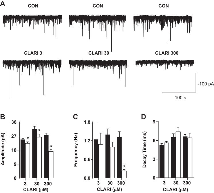
Fig. 3.
Clarithromycin inhibits miniature GABAergic currents. A: representative traces of mIPSC recordings from 3 representative neurons bathed in ACSF containing CNQX, d-AP5, and TTX before (top) and after (bottom) application of clarithromycin. B–D: bar graph pairs represent average values ± SE in controls (filled bars) and in 3, 30, and 300 μM clarithromycin (open bars). B: mIPSC amplitude was significantly reduced in the presence of all tested concentrations of clarithromycin (CON vs. CLARI 3: 24.8 ± 0.6 pA vs. 22.3 ± 0.9 pA, n = 15; CON vs. CLARI 30: 31.5 ± 1.9 pA vs. 26.4 ± 1.9 pA, n = 12; CON vs. CLARI 300: 27.6 ± 1.2 pA vs. 17.1 ± 1.1 pA, n = 9; paired t-test, *P = 0.02, *P = 0.001, *P = 0.0001, respectively). C: the frequency of current events was almost completely abolished at high dose (CON vs. CLARI 300: 1.3 ± 0.5 Hz vs. 0.23 ± 0.14 Hz; n = 9; paired t-test, *P = 0.0001). D: decay time was not changed with clarithromycin application (P > 0.1).

