
Fig. 1.
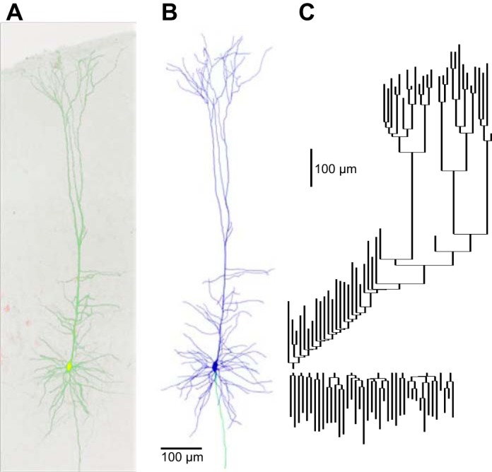
Morphological reconstruction of the representative corticospinal neuron. A: 2-photon fluorescence image (maximum intensity projection) of a corticospinal neuron filled intracellularly with fluorescent dye (green); the red channel shows numerous corticospinal neurons retrogradely labeled with fluorescent beads (red). B: 3-dimensional reconstruction of the same neuron. Blue, dendrites; green, axon. C: dendrogram representation of the dendritic arbor topology of the reconstructed neuron. Apical branches point upwards and basal branches point downward. Vertical distance represents dendritic path length; horizontal connectors are dimensionless.