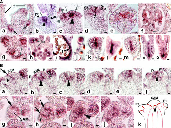
Figure 3.
HAN Expression Pattern.
(A) Budding axillary SAMs are shown in (a). Arrows point to the expression domains of HAN at the boundaries between the SAM and the adaxial sides of leaves. Lf, leaf. (b) Older axillary SAM. Arrows point to the expression domains of HAN at the boundaries between the SAM and the newly initiated organ primordia (pr, small arrowheads). Large arrowhead indicates the junction of the HAN expression domain in the SAM and its vascular expression in the stem. (c) Inflorescence SAM. Arrows point to two stripes of HAN expression at the boundaries between the SAM and newly initiated floral primordia. Small arrowheads indicate HAN expression in a stage 2 flower at the boundaries between the floral meristem and the soon-to-be-specified sepal primordia. Large arrowheads point to the junction of the HAN expression domain in the SAM and in the stem (note that the angle of this section is tilted toward the reader, and the domain indicated by the large arrow includes some of the expression of HAN between the SAM and a primordium pointing toward the reader). (d) Stage 5 flower. Arrow points to the connection arch of HAN expression in the floral meristem and its expression in the peduncle vascular tissue. Se, sepal. (e) Expression in the lateral and basal regions of carpel primordia in a stage 6 flower. (f) Early stage 12 ovary (cross section). HAN is expressed in initiating inner and outer integuments as well as in the vascular tissue (arrows). (g) Expression in integuments continues in the stage 13 ovary. Arrow points to expression in funiculus vascular tissue. (h) HAN expression in the stamens of a stage 8 flower. lo, locule. (i) HAN expression persists in the tapetum cell layer until it has degenerated and is absent in mature haploid pollen. t, tapetum. (j) Eight-cell stage embryo. HAN is expressed in all cells of the embryo proper. (k and l) Globular and transition stage embryos. HAN is expressed in the center files of cells. (m and n) Heart and torpedo stage embryos. HAN expression remains in the center cells destined to be provascular tissues. (o) Late torpedo stage. Bars = 10 μm.
(B) Expression in SAM in serial longitudinal (a to f) and cross sections (g to j). Arrows point to expression at the boundaries between the SAM and new organ primordia as well as between organ primordia. Expression of HAN in the SAM merges with its expression in the vascular strands in the stem (arrowheads) as illustrated in (k). In (k), red lines represent the expression domains of HAN in the SAM, stem, and developing organ primordia (P1 and P2). Arrowhead corresponds to the regions indicated by large arrowheads in Figure 3A (b and c). P, primordium. Bars = 10 μm.

