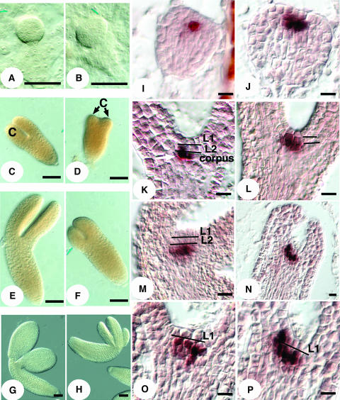
Figure 6.
han Mutant Embryo Defects and Perturbed WUS Expression in han Embryos.
(A) Globular stage wild-type embryo.
(B) Globular stage han-1 mutant embryo.
(C) Late heart stage wild-type embryo. C, cotyledon.
(D) Late heart stage han-1 embryo with stunted cotyledons.
(E) Walking stick stage wild-type embryo.
(F) Walking stick stage han-1 embryo.
(G) Mature wild-type embryo.
(H) Mature han-1 mutant embryo with three cotyledons.
(I) WUS expression in the transition stage wild-type embryo is concentrated in two cells in the subepidermal L2 layer.
(J) WUS expression in the transition stage han-1 embryo is located in more than two cells within and beneath the L2 layer.
(K) WUS expression in the heart stage wild-type embryo is shifted to two central cells in the corpus.
(L) WUS expression in the heart stage han-1 embryo in the L2 layer.
(M) WUS expression in the mature stage wild-type embryo is centered below the outermost two layers.
(N) WUS expression in the mature han-1 embryo.
(O) and (P) WUS expression in the L2 layer of mature han-1 embryos. (P) is an enlarged view of (N).
Bars = 50 μm in (A) to (H) and 10 μm in (I) to (P).

