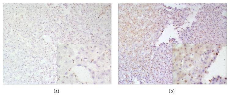
Figure 7.
Ki-67 staining of proliferating hepatocytes in liver of the experimental monkey. (a) Preoperative liver section showed a negative expression. (b) Liver section from postoperative day 10 revealed a few positively stained nuclei (×10). High magnification (×20) images are shown in the lower right corner.

