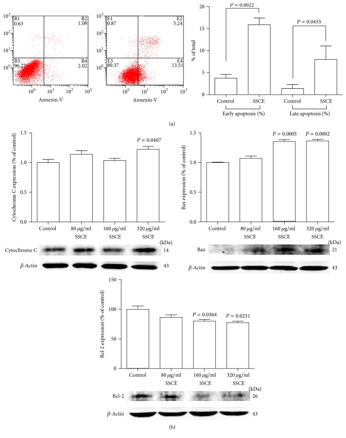
Figure 2.
(a) SSCE (160 μg/ml) treatment for 24 h induced apoptosis of MCF-7 cells with a significant increase in the proportion of both early and late apoptotic cells analyzed by FACS. (b) Western blot analysis and quantification of the band intensity show that after SSCE (80 μg/ml, 160 μg/ml, and 320 μg/ml) treatment for 24 h, the expression levels of apoptosis-related proteins cytochrome C and Bax were elevated, and Bcl-2 expression was suppressed.

