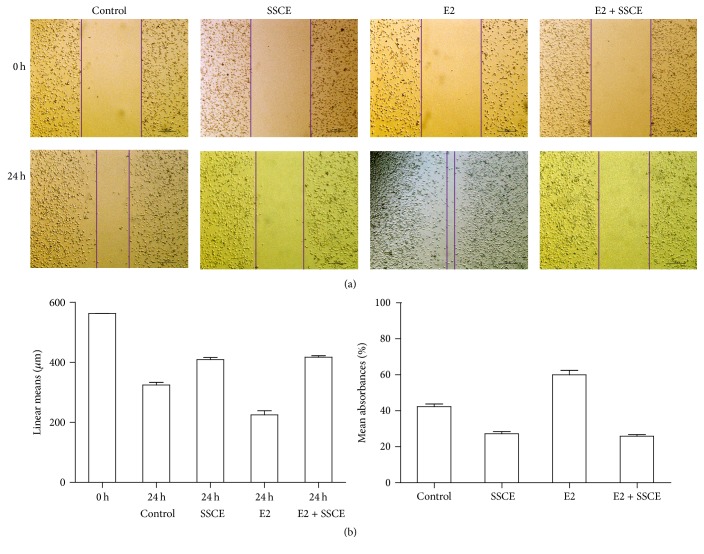
Figure 4.
Wound healing assays to assess the effect of SSCE on migration ability. MCF-7 cells were cultured in a 6-well plate to approximately 90% confluency. A wound was generated by a scratch in the middle of the plate using a pipette. The wound closure was measured upon treatment with E2 (10−8 M) and SSCE (160 mg/ml) alone or E2 (10−8 M) + SSCE (160 mg/ml) for 24 h. (a) Representative images show the wound at 0 and 24 h. The migration of MCF-7 cells was promoted by E2 and inhibited by SSCE both in the presence and absence of E2. (b) The linear mean length and the confidence interval for each wounded area 24 h after the scratch are presented and compared to 0 h. The linear mean length of the control and the cells treated with SSCE and SSCE + E2 were significantly reduced compared to that in the other groups (P < 0.001). There was a significant difference noted in the linear means between the control and the E2-treated cells (P < 0.001). The rate of closure with the condition interval for each group 24 h after scratch is presented. The rates of closure of the SSCE group and SSCE + E2 group were significantly lower than that in the other groups.

