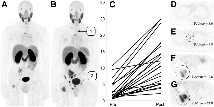
FIGURE 2.
(A and B) Coronal maximum-intensity projections of patient with castration-sensitive metastatic prostate cancer imaged using 68Ga-PSMA-11 before ADT (A) and after ADT (B) demonstrate marked increase in uptake in lesions. (C) Each visualized lesion demonstrated increased uptake, averaging more than 7 times the initial uptake. (D and E) Numerous lesions (13 of 22) were visualized only on posttreatment imaging, as exemplified by the upper thoracic osseous metastasis seen on these axial PET images. (F and G) Other lesions increased in size and had increased uptake on posttreatment imaging, as exemplified by the lesion seen on these axial PET images.

