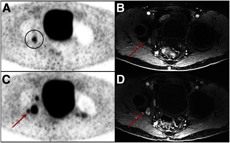
FIGURE 3.
Examples of lesions seen only on post-ADT PSMA PET. (A) Pretreatment PET image demonstrates single lesion (circled) in right acetabulum. (B) Additional non–PSMA-avid right acetabular lesion (arrow) is seen just lateral to larger lesion on MR image of same location. (C) On post-ADT PET image, numerous additional lesions are seen, including adjacent acetabular lesion (arrow). (D) This lesion (arrow) is again demonstrated on enhanced T1-weighted MR image.

