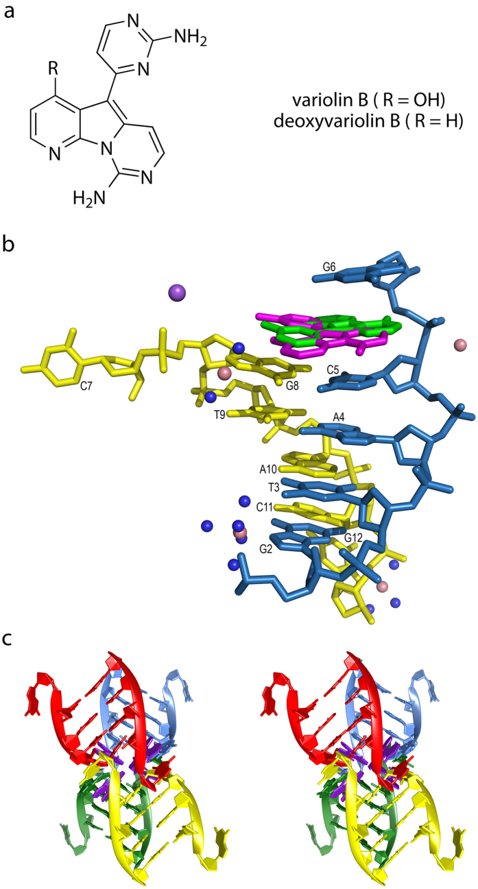
Figure 1.
(a) Chemical structure of variolin B and the deoxyvariolin B derivative. (b) Content of the asymmetric unit of the d(CGTACG)2-variolin B crystal. Both half-occupancy drug molecules are shown. Cobalt and sodium atoms are represented with pink and purple spheres, respectively. Water molecules are not shown for clarity. (c) Stereoview of the four interlaced DNA duplexes (red, blue, green and yellow) forming four intercalation sites. One variolin B molecule is shown in purple in each of these sites.

