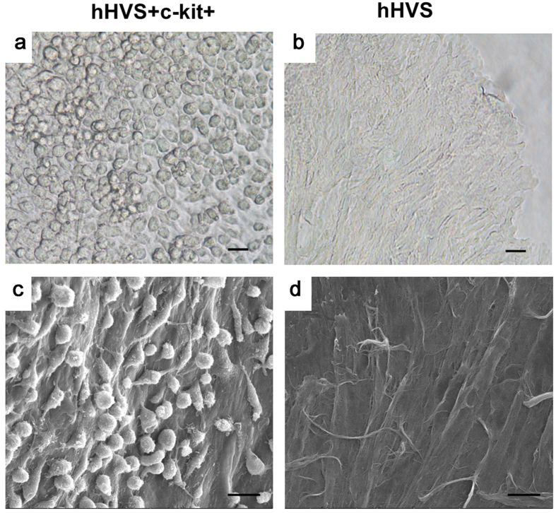
Figure 2. Cell-scaffold adhesion.
(a) Adhesion of growing BM c-kit+ cells on the hHVS under optical microscope after 10 days in cell culture. (b) hHVS without growing cells under the optical microscope. (c) Adhesion of growing BM c-kit+ cells on the hHVS under scanning electron microscope after 10 days cell culture. (d) hHVS without growing cells under the scanning electron microscope. Scale bar, 20 μm. Data shown are representative of 4 independent experiments.

