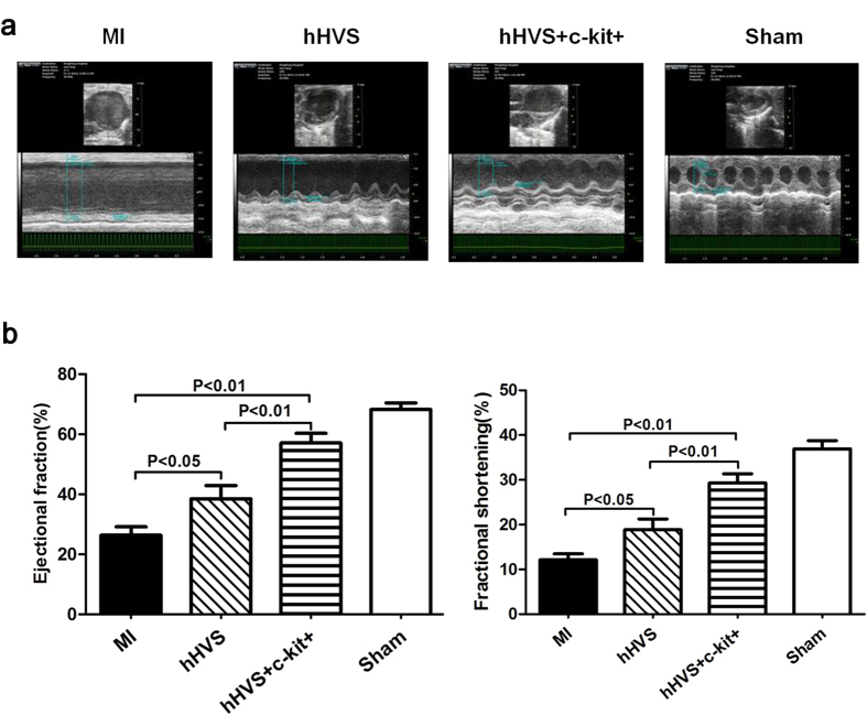
Figure 5. Echocardiographic examination 4 weeks after MI/implantation.
(a) Representative echocardiography of MI (without patch implantation), hHVS (MI with implantation of hHVS alone), hHVS + c-kit+ (MI with implantation of c-kit+ cell-seeded hHVS), and sham groups. (b) Comparisons of ejection fraction (EF), fractional shortening (FS). MI, n = 10; hHVS, n = 10; hHVS + c-kit+, n = 10; sham, n = 7.

