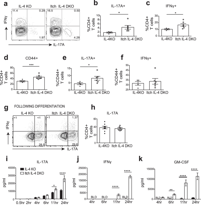
Figure 5. Itch E3 ligase is required to restrict numbers of Th17 cells and secretion of proinflammatory Th17 cytokines.
(a–f) Flow cytometric analysis of lung cells from Itch IL-4 DKO mice or IL-4KO controls. (a) Percentage of IL-17A+ or IFNγ+ CD4+ cells. (b,c) Percentage of IL-17A+ (b) or IFNγ+ (c) among CD3+ CD4+ lung cells. (d–f) Percentages of previously activated CD44+ lung cells among CD4+ T cells (d) and percentages of IL-17A+ (e) or IFNγ+ (f) previously activated cells shown in panel D. (g–k) Naïve sorted CD4+ T cells were differentiated into Th17 cells for 5 days and expanded in IL-2. (g) Representative plot showing IL-17A+ or IFNγ+ Th17 cells after differentiation. (h) Summary of data over multiple experiments. (I,k) Cytokines were analyzed using ELISA following restimulation of equal numbers of IL-2 expanded Th17 cells. (i) IL-17A (j) IFNγ and (k) GM-CSF. In all summarized plots, each dot represents an individual mouse analyzed in at least 2 independent experiments. Significance was calculated by unpaired T tests for panel b–f, h and by 2-way ANOVA for panels i-k. *p < 0.05, **p < 0.01,***p < 0.001, ****p < 0.0001. All error bars represent mean +/− SEM.

