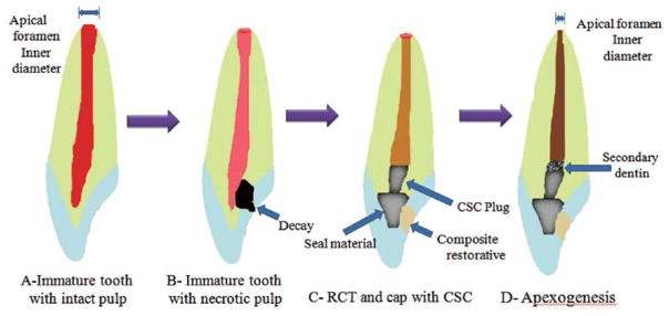
Fig. 3. Schematic presentation of dental pulp regeneration processes in an immature tooth.
A: Immature tooth with intact pulp; B: Necrotic dental pulp and accumulation of stem cells of the apical papilla (SCAP) in periapical area; C: Mechanical provocation of bleeding from the periapical site to transport the SCAPs into the empty canal space and establishment of a coronal seal by MTA cement and composite resin materials; D: Formation of pulp-like tissue in apical and mid third of root, which contributes to the maturation of apical third and apical foramen closure by the formation of a cementum-like tissue.

