
Fig. 2.
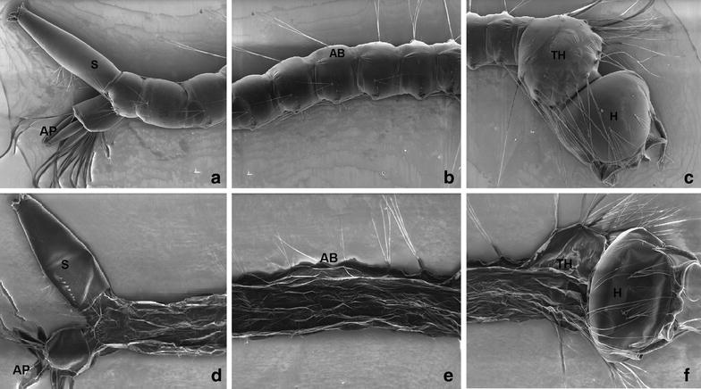
Scanning electron micrograph of C. quinquefasciatus larvae. Control (a–c) showing no alteration on head (H), thorax (TH), abdomen segments (AB), siphon (S) and anal papillae (AP). Larvae treated with P. emarginatusnanoemulsion at 250 ppm (d–f) showing alterations on cuticle of abdomen (AB), thorax (TH) and anal papillae (AP)