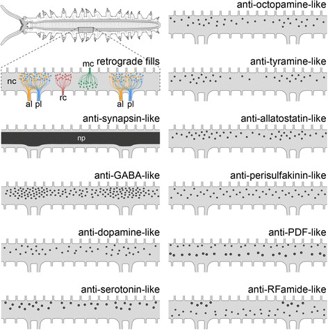
Fig. 10.

Simplified diagrams of onychophoran nerve cords summarising the results of retrograde fills and immunohistochemistry. The upper left diagram shows the entire central nervous system in dorsal view (anterior is left). The remaining diagrams illustrate detailed nerve cords from one body side of two consecutive leg-bearing regions. al, anterior leg nerve; mc, median commissure; nc, nerve cord; pl, posterior leg nerve; rc, ring commissure
