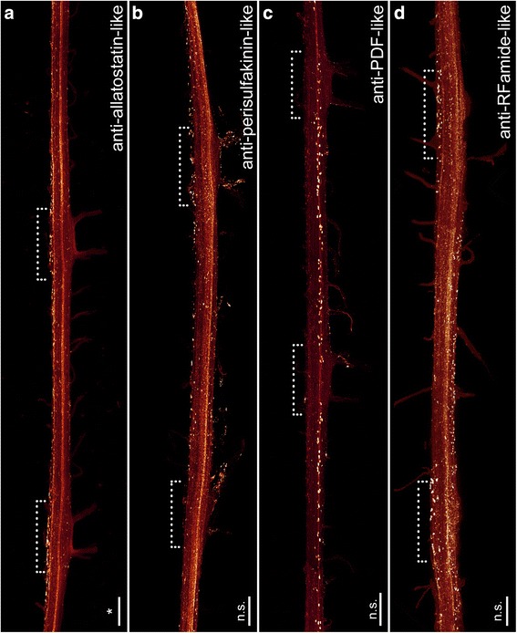
Fig. 7.

Organisation of individual ventral nerve cords in Onychophora as revealed by different neuronal markers: antisera against allatostatin (a), perisulfakinin (b), pigment-dispersing-factor (PDF; c) and RFamide (d). Maximum projection confocal micrographs of Euperipatoides rowelli. Median is left. Asterisks indicate the distribution of somata that is significantly different from uniform (χ2 *, **, ***, ****, p < 0.05, 0.01, 0.001, 0.0001 respectively; n.s. not significant; see Table 1 for more details). Dotted brackets indicate leg-bearing regions characterised by the position of leg nerves. d Note the condensed groups of neuronal cell bodies labelled against the peptide RFamide that are present near the bases of leg nerves. Scale bars: 200 μm
