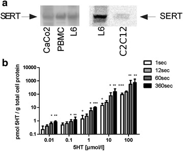
Fig. 1.

a SERT-expression in L6 and C2C12 skeletal muscle cells. Western blot analysis of total cell lysates using a SERT-specific antibody. Expression of SERT was compared between CaCo2 cells (human colon carcinoma cell line) and PBMC cells (monocyte cell line) two cell lines for which SERT-expression has been previously described [16, 17], and L6 cells (N = 6) and C2C12 cells respectively (N = 2). b Concentration- and time-dependent uptake of 5-HT into L6 cells using radioactive 5-HT. L6 cells were incubated for increasing incubation times (1, 12, 60 and 360 s) and increasing [3H]-labelled 5-HT concentrations (0.01; 0.1; 1; 10 and 100 µM) and analysed for 5-HT uptake. Bars represent the mean ± S.D. for three experiments performed in triplicates. Significant differences are indicated as *P < 0.05 **P < 0.01 ***P < 0.001 for comparisons to the condition at 1 s incubation time and at the same 5-HT concentration, and +P < 0.05 +++P < 0.001 for comparisons to condition at 1 s incubation time and at the 5-HT concentration of 0.01 µM
