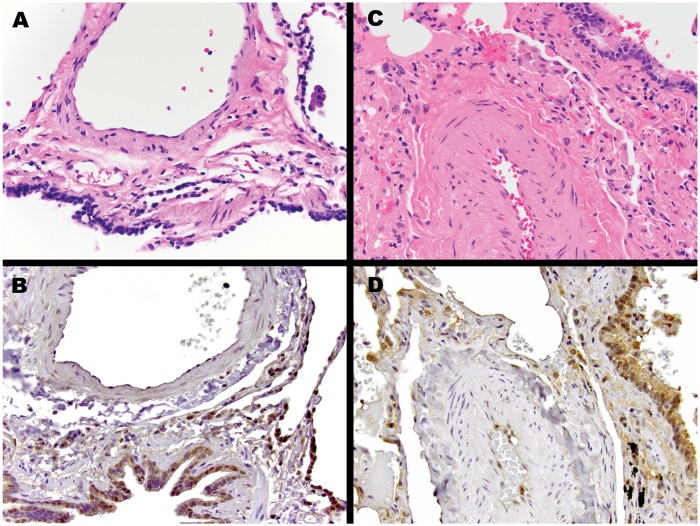
Figure 9.
Macrophage migration inhibitory factor (MIF) immunohistochemistry. Representative pulmonary parenchyma from control patient without pulmonary arterial hypertension (PAH) or lung disease (A and B) and patient with PAH (C and D). Compared with the control lung, the pulmonary artery from the patient with PAH demonstrates thickening and hypertrophy of the muscular medial layer. MIF immunostaining for both patients with PAH and control patients show expression in airway-lining ciliated bronchial epithelial cells, pulmonary macrophages, pneumocytes, and endothelial cells. Of note, the vascular smooth-muscle cells are negative. Hematoxylin and eosin stain (A, C) and MIF immunostain (B, D) images are presented at 400× original magnification.

