
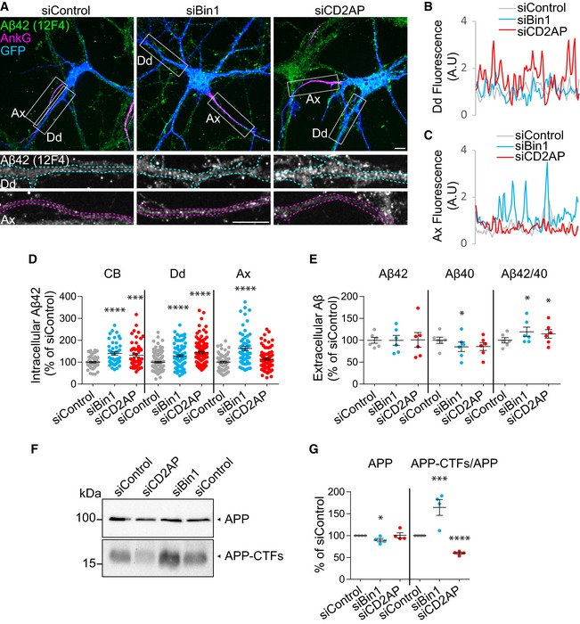
Figure 1. Downregulation of Bin1 and CD2AP increases polarised endogenous Aβ generation.
-
AIntracellular endogenous Aβ42 (green), Ankyrin‐G (AnkG; magenta) and GFP (blue) in siBin1‐, siCD2AP‐ and siControl‐treated primary cortical neurons (neurons) expressing GFP immunolabelled at 9 DIV with anti‐Aβ42 (clone 12F4) and anti‐AnkG, analysed by spinning‐disc confocal microscopy. The white rectangles indicate the dendrites (Dd) and axons (Ax) magnified below showing Aβ42 in axons and dendrites outlined based on AnkG (magenta) and GFP (blue), respectively. Scale bars, 10 μm.
-
B, CAβ42 line profiles in dendrites (Dd; B) and axons (Ax; C) of siControl (grey line), siBin1 (blue line) and siCD2AP (red line) neurons shown in (A).
-
DQuantification of Aβ42 (12F4) intensity in cell body (CB), dendrite (Dd) and axon (Ax) (n = 5, N CB = 44–53, N Dd = 90–120, N Ax = 60–74; ****P CB < 0.0001 siBin1 vs. siControl, ***P CB < 0.001 siCD2AP vs. siControl, ****P Dd < 0.0001 siBin1 vs. siControl, ****P Dd < 0.0001 siCD2AP vs. siControl, ****P Ax < 0.0001 siBin1 vs. siControl, t‐test, mean ± SEM).
-
EQuantification of extracellular endogenous Aβ40, Aβ42 and of Aβ42/Aβ40 ratio by ELISA analysis of conditioned media of 9 DIV siBin1, siCD2AP or siControl neurons (n = 6; *P Aβ40 = 0.0270 siBin1 vs. siControl, *P Aβ42/40 = 0.0378 siBin1 vs. siControl, *P Aβ42/40 = 0.0463 siCD2AP vs. siControl, t‐test, mean ± SEM).
-
FEndogenous APP and APP‐CTFs levels by Western blot with anti‐APP antibody (Y188) of siBin1‐, siCD2AP‐ or siControl‐treated neurons at 9 DIV.
-
GQuantification of APP and APP‐CTFs levels normalised to APP (n = 4; *P APP = 0.0355 siBin1 vs. siControl, ***P APP‐CTFs/APP < 0.001 siBin1 vs. siControl, ****P APP‐CTFs/APP < 0.0001 siCD2AP vs. siControl, t‐test, mean ± SEM).
Source data are available online for this figure.