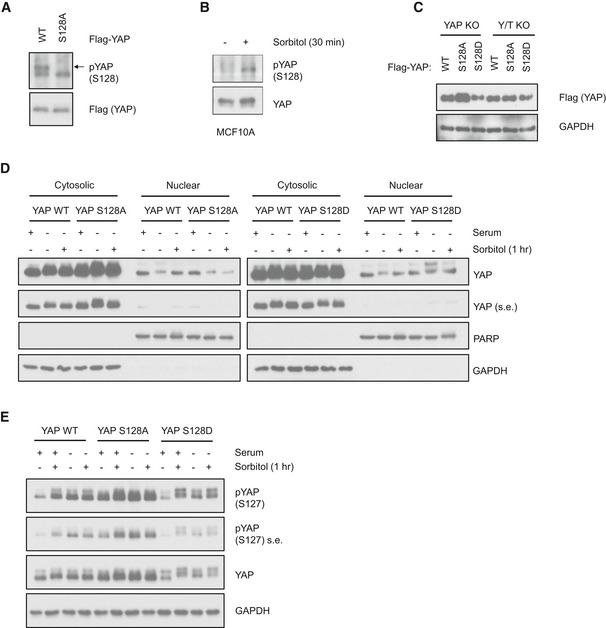
Figure EV4. Osmotic stress induces YAP Ser128 phosphorylation to determine its subcellular localization.

- pYAP S128 antibody is specific to YAP S128 site. Flag‐YAP wild‐type (WT) and S128A mutant constructs were transfected into HEK293A cells. Cell lysates were collected for Western blot analysis by indicated antibodies.
- Osmotic stress induces endogenous YAP Ser128 phosphorylation in MCF10A cells. MCF10A cells were treated with sorbitol and endogenous YAP was immunoprecipitated. YAP Ser128 phosphorylation and protein levels were determined by Western blot.
- Expressions of Flag‐YAP WT, S128A, and S128D mutant stable cell lines are at similar levels. Stable cell lines were generated with retroviral infection of YAP WT and mutant constructs into YAP KO or YAP/TAZ dKO HEK293A cells. Cell lysates were collected for Western blot analysis. YAP expression level was detected by Flag antibody, with GAPDH as a loading control.
- Subcellular fractionation of YAP WT‐, S128D‐, and S128A‐reconstituted cells. YAP KO HEK293A cells were stably reconstituted with YAP WT, S128D, or S128A. Cytosolic and nuclear fractions were collected by differential fractionation. PARP and GAPDH were used as nuclear and cytosolic markers, respectively. s.e. denotes short exposure of the YAP Western blot.
- Osmotic stress induces YAP Ser127 phosphorylation despite Ser128 phosphorylation status. YAP KO HEK293A cells were stably reconstituted with YAP WT, S128D, or S128A mutants and were treated with sorbitol in the presence or absence of serum. YAP Ser127 phosphorylation and protein levels were determined by Western blot. s.e. denotes short exposure.
Source data are available online for this figure.
