Figure 1.

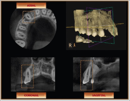
Cone beam computed tomography images of maxillary right arch obtained for the study subjects are shown in three-dimensional, axial, coronal, and sagittal views

Cone beam computed tomography images of maxillary right arch obtained for the study subjects are shown in three-dimensional, axial, coronal, and sagittal views