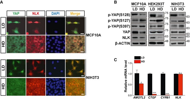
Level of endogenous NLK mRNA is unaffected, whereas the expression of YAP target genes was reduced, at high cell density. Quantitative real‐time PCR analyses for expression of AMOTL2, CTGF, CYR61, and NLK in low‐density (LD) and high‐density (HD) NIH3T3 cells were performed. Quantification of AMOTL2, CTGF, CYR61, and NLK mRNA was normalized with the level of Gapdh. Data represent average values from a representative of multiple experiments performed in triplicate. Error bars indicate standard deviations of triplicate measurements. Data are presented as mean ± SD. *P < 0.05 and ***P < 0.005. Student's t‐test was used for statistical analysis.