-
A
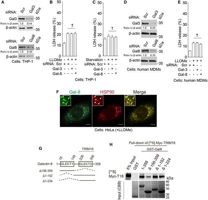
Immunoblot analyses of knockdown efficacies in THP‐1 cells.
-
B, C
LDH release data for samples in (B) Fig
2A and (B) Fig
2B.
-
D
Immunoblot analyses of knockdown efficacies in primary human MDMs.
-
E
LDH release data for samples in Fig
2C.
-
F
Confocal microscopy of HeLa cells treated with LLOMe and stained for galectin‐8 and HSP90. Arrowheads in enlarged insets indicate colocalization. Scale bars, 5 μm.
-
G
Galectin‐8 domains and deletion constructs used.
-
H
In vitro translated and radiolabeled [35S] myc‐HA‐TRIM16 was incubated with full‐length‐ and deletions of GST‐galectin‐8 in the presence of flag‐ULK1 and cold ATP, and GST pull downs were performed and [35S] radiolabeled Myc‐HA‐TRIM16 detected by SDS‐PAGE and autoradiography. Amounts of GST fusion proteins are shown in Coomassie brilliant blue (CBB)‐stained gels.
Data information: Means ± SEM;
n ≥ 5, except for immunoblot quantifications where
n ≥ 3.
†
P ≥
0.05 (ANOVA for B, C, E).