
Figure EV3. Disease‐associated PEX13 mutant protein colocalization with peroxisomes and effects on mitochondrial morphology, and structured illumination microscopic analyses of mitochondria in PEX13 patient mutant fibroblasts.
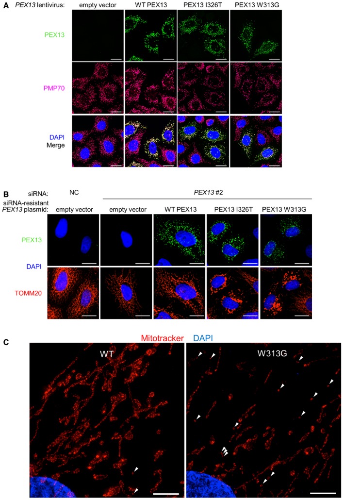
- Representative images of wild‐type and mutant PEX13 colocalization with PMP70 in HeLa/Parkin cells transduced with lentivirus containing the indicated PEX13‐MYC‐DDK cDNA. Scale bars, 20 μm.
- Representative images of TOMM20 morphology 16 h after DMSO vehicle treatment in HeLa/Parkin cells transfected with PEX13 siRNA and indicated PEX13 siRNA‐resistant plasmids. Scale bars, 20 μm.
- Representative structured illumination microscopy (SIM) 3D projections of wild‐type and PEX13 W313G mutant primary human fibroblasts treated with MitoTracker CMX rosamine (CMXROS, 50 nM, 30 min). White arrowheads indicate mitochondrial fragments. Scale bar, 5 μm.