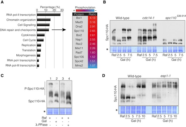
Figure 5. The spindle pole body component Spc110 is a target of Cdc14 during the DNA damage response.

- Identification of Cdc14 phospho‐targets during the DNA damage response by mass spectrometry analysis. Wild‐type and cdc14‐1 cells were grown overnight and blocked in G2/M by using nocodazole to avoid cell cycle‐dependent changes in protein phosphorylation between both strains. After the block was attained, cells were transferred to 37°C for 45 min to deplete Cdc14 activity prior to HO induction by galactose addition for 4 h. Differential phosphorylation of phospho‐peptides detected between the wild‐type and cdc14‐1 were grouped into broad categories depending on the molecular function of the proteins. The table includes the DNA damage and checkpoint‐related proteins with Cdc14‐dependent hyper‐phosphorylated status and the relative ratio between the wild‐type and cdc14‐1 during the DNA damage response. Red and blue indicate relative amount of the residue phosphorylation between both strains (red, high; blue, low).
- Cdc14 dephosphorylates Spc110 in response to a DSB. Spc110 was tagged with the 6HA epitope in wild‐type, cdc14‐1 and spc110 S36‐91A backgrounds. Cells were grown overnight in raffinose‐containing media and supplemented with galactose to induce HO expression. Samples were collected at each time point indicated. Proteins were TCA extracted, separated on Phos‐Tag‐containing gels and blotted. Coomassie staining is shown as a loading control.
- Lambda‐phosphatase treatment of protein extracts in the absence of DNA damage reduces Spc110 phosphorylation levels to the same extent as samples taken during the DNA damage response. Cells were grown overnight in raffinose‐containing media (line 1) and incubated with galactose for 2.5 h to induce HO expression (line 2). Extracts from raffinose‐containing cultures were treated with mock (line 3) or λ‐PPase (line 4). All samples were separated in a Phos‐Tag‐containing gel and blotted. Coomassie staining is shown as a loading control.
- The canonical FEAR pathway is required for Spc110 dephosphorylation during the DNA damage response. Spc110 was labelled with the 3HA tag in both wild‐type and esp1‐1 background strains. Cells were grown overnight in raffinose‐containing media and supplemented with galactose to induce the HO expression. Samples were collected at each time point indicated. Proteins were TCA extracted, separated on Phos‐Tag‐containing gels and blotted. Coomassie staining is shown as a control for loading.
