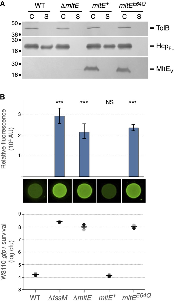
Anti‐bacterial activity. Escherichia coli K‐12 prey cells (W3110 gfp
+, kanR) were mixed with the indicated attacker cells, spotted onto Sci‐1 inducing medium (SIM) agar plates, and incubated for 4 h at 37°C. The image of a representative bacterial spot and the average and standard deviation (n = 3) of the relative fluorescence of the bacterial mixture (in arbitrary units, AU) are shown in the upper graph. The number of recovered E. coli prey cells (counted on selective kanamycin medium) is indicated in the lower graph [in log10 of colony‐forming units (cfu)]. The black, dark gray, and light gray circles indicate values from three independent assays, and the average is indicated by the bar. The experiment was performed in triplicate and a representative result is shown. Asterisks indicate significant differences compared to the wild‐type attacker strain (NS, non‐significant; ***P < 0.001; Student's t‐test).

