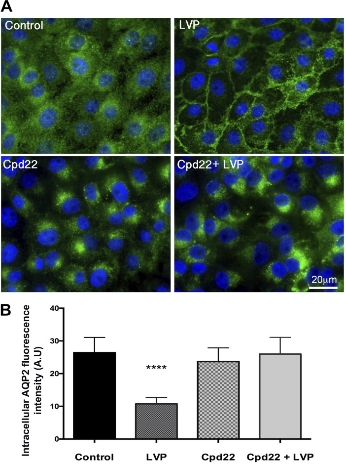
Fig. 2.
A: immunofluorescence staining showing the baseline distribution of AQP2 in control cells and the accumulation of AQP2 in the plasma membrane after 15 min of vasopressin treatment. ILK inhibition with Cpd22 blocked vasopressin-induced membrane accumulation of AQP2 and a perinuclear patch of AQP2 developed in these cells. Scale = 20 μm. Green, AQP2; blue, nucleus. B: intracellular AQP2 fluorescence intensity arbitrary units (AU), showing a significant decrease in intracellular AQP2 in LLC-AQP2 cells treated with vasopressin (LVP) but not in LLC-AQP2 cells treated with vasopressin after Cpd22 treatment. Bar values represent ± SE. ****P < 0.0001, n ≥ 3. Statistics were done by 1-way ANOVA. The mean of each column was compared with control mean.

