Fig. 3.
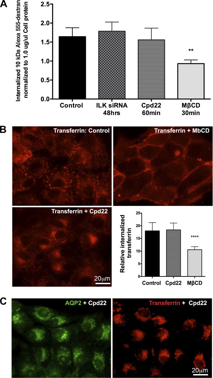
A: a bar graph showing fluid phase endocytosis assay results of internalized fluorescence of 10 kDa Alexa 555-dextran. There was no significant difference in endocytosis of 10 kDa Alexa 555-dextran in LLC-AQP2 cells after ILK inhibition by both siRNA and cpd22. A block of endocytosis with methyl-β-cyclodextrin (MβCD) was used as a positive control and significantly inhibited 10 kDa Alexa 555-dextran endocytosis compared with any of the treatments. B: the clathrin endocytosis pathway was assessed by a rhodamine-conjugated transferrin endocytosis assay, which showed accumulation of transferrin at the perinuclear region in cells treated with ILK inhibitor cpd22 when compared with controls. However, quantification of the endocytosed transferrin revealed no difference in the overall endocytosis rate between controls and ILK inhibited cells. A block of endocytosis with MβCD was used as a positive control. C: double staining showing colocalization of AQP and endocytosed transferrin in the perinuclear patch after ILK inhibition, suggesting that clathrin-endocytosed cargo is accumulated at the perinuclear region upon ILK inhibition. Scale = 20 μm. Green, AQP2; red, transferrin. Bar values represent ± SE. **P < 0.01, **** P < 0.0001, n ≥ 3. Statistics were done by 1-way ANOVA. The mean of each column was compared with control mean.

