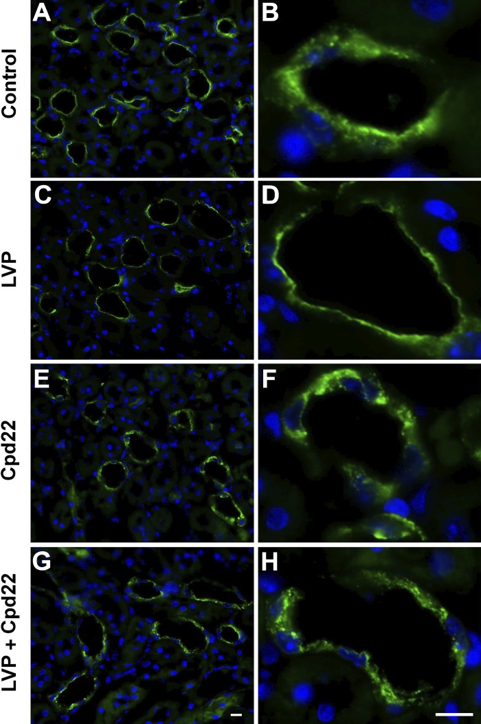
Fig. 11.
Immunofluorescence images showing cpd22 inhibits vasopressin-associated AQP2 plasma membrane accumulation in collecting ducts from wild-type mice kidney slices in vitro. A, B: control tissues treated with HBSS alone for 75 min. C, D: 45 min treatment of HBSS alone followed by 30 min AVP treatment, which caused accumulation of AQP2 on the apical plasma membrane. E, F: 75 min of cpd22 in HBSS treatment showing patches of AQP2 accumulation within principal cells. G, H: 30 min AVP in HBSS treatment after 45 min cpd22 treatment showing no accumulation of AQP2 in the membrane and the presence of large intracellular patches of AQP2. Scale = 15 µm. Green, AQP2; blue, nucleus.

