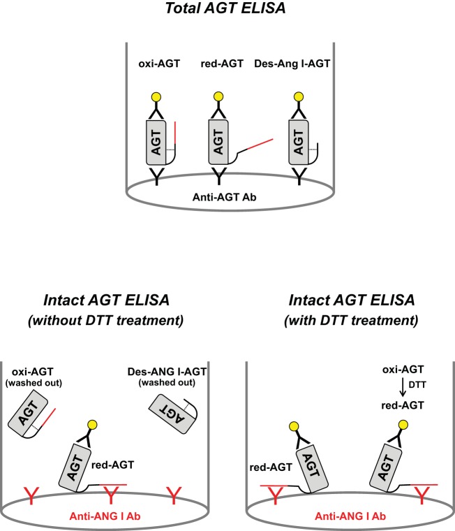
Fig. 1.
Figure depicting the system of total angiotensinogen (tAGT) ELISA and intact AGT (iAGT) ELISA. Red line in AGT molecule indicates ANG I region. Dotted line shows a disulfide bond in AGT molecule. In tAGT ELISA, two different anti-AGT antibodies are used. In iAGT ELISA, a combination of an anti-ANG I antibody and an anti-AGT antibody was used. iAGT ELISA without dithiothreitol (DTT) treatment detects only endogenous reduced intact AGT (red-iAGT).

