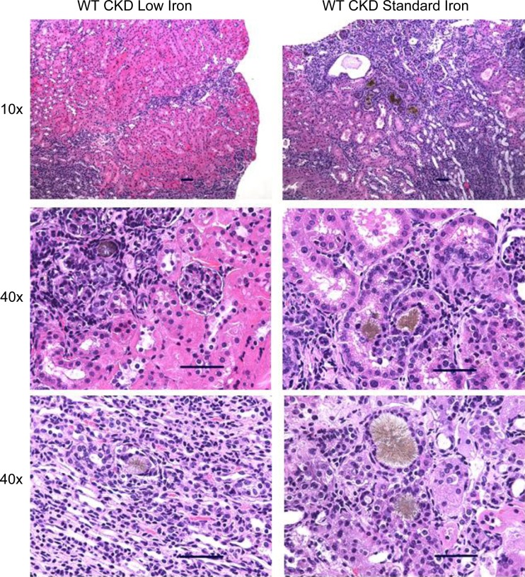
Fig. 5.
Renal histopathological analysis of mice on the adenine diets. Kidney tissue sections were stained with hematoxylin and eosin and assessed with light microscopy at ×10 and ×40 magnification. The adenine diet induced significant peritubular leukocyte infiltration, deposition of crystalloid structures in the tubular lumina, and tubular dilatation. Scale bar = 50 μm.

