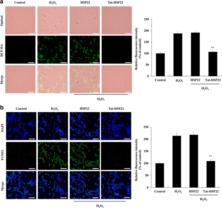
Fig. 4.

The effect of Tat-HSP22 protein against H2O2-induced intracellular toxicities in HT-22 cells. HT-22 cells were pretreated with 5 μM of Tat-HSP22 or control HSP22 protein for 2 h and the cells were incubated in the absence or presence of H2O2 (1 mM) for 10 min and 6 h, respectively. Then, a H2O2-induced oxidative stress levels were measured using DCF-DA staining and the fluorescence intensity was measured using an ELISA plate reader. b DNA fragmentation was detected by TUNEL staining and quantitative evaluation of TUNEL positive cells confirmed by cell counting under a phase-contrast microscopy. Scale bar = 50 μm. ** P < 0.01 compared with H2O2-treated cells
